
Sign in
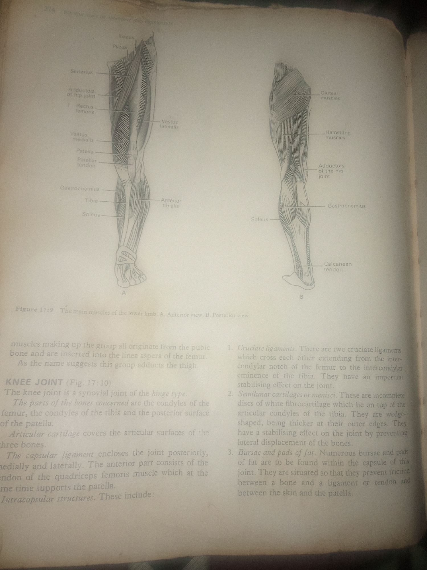
This is a photograph of an open book, showing an anatomical illustration of the human leg muscles and a detailed description of the knee joint. The illustration is divided into two views: A, the anterior view of the lower limb muscles, and B, the posterior view. Various muscles are labeled, including the sartorius, rectus femoris, vastus medialis, vastus lateralis, gastrocnemius, soleus, and patellar tendon. The text describes the muscles making up a group that originates from the pubic bone and inserts into the femur, adducting the thigh. It then delves into the anatomy of the knee joint, identifying it as a synovial hinge type. Key components like the condyles of the femur and tibia, the patella, articular cartilage, and capsular ligament are mentioned. The text also elaborates on intracapsular structures, specifically cruciate ligaments, semilunar cartilages (menisci), and bursae and fat pads, detailing their functions in stabilizing the joint and preventing friction. The image is a clear, black and white illustration, typical of an anatomy textbook. The page appears to be slightly yellowed, suggesting it is an older publication. No information about time of day, weather, or specific emotions is available as the image focuses on scientific illustration and text. The location context provided (Jalingo, Nigeria) is external to the image content itself.
FM-KpyEx1

Jun 30, 2026, 4:14 PM

Jalingo, Nigeria

Stake attention in this memory

educational
informative
anatomical
scientific
detailed

This is a photograph of an open book, showing an anatomical illustration of the human leg muscles and a detailed description of the knee joint. The illustration is divided into two views: A, the anterior view of the lower limb muscles, and B, the posterior view. Various muscles are labeled, including the sartorius, rectus femoris, vastus medialis, vastus lateralis, gastrocnemius, soleus, and patellar tendon. The text describes the muscles making up a group that originates from the pubic bone and inserts into the femur, adducting the thigh. It then delves into the anatomy of the knee joint, identifying it as a synovial hinge type. Key components like the condyles of the femur and tibia, the patella, articular cartilage, and capsular ligament are mentioned. The text also elaborates on intracapsular structures, specifically cruciate ligaments, semilunar cartilages (menisci), and bursae and fat pads, detailing their functions in stabilizing the joint and preventing friction. The image is a clear, black and white illustration, typical of an anatomy textbook. The page appears to be slightly yellowed, suggesting it is an older publication. No information about time of day, weather, or specific emotions is available as the image focuses on scientific illustration and text. The location context provided (Jalingo, Nigeria) is external to the image content itself.

Loading AttnAds…

transactions
revenues
stakers
Earliest
Latest
Highest stake

No transactions found

More from this user

More from Jalingo

FM-KpyEx1

Jun 30, 2026, 4:14 PM

Jalingo, Nigeria

Licensed under Creative Commons Attribution 4.0 International License. You are free to share and adapt this content with proper attribution.