
Sign in
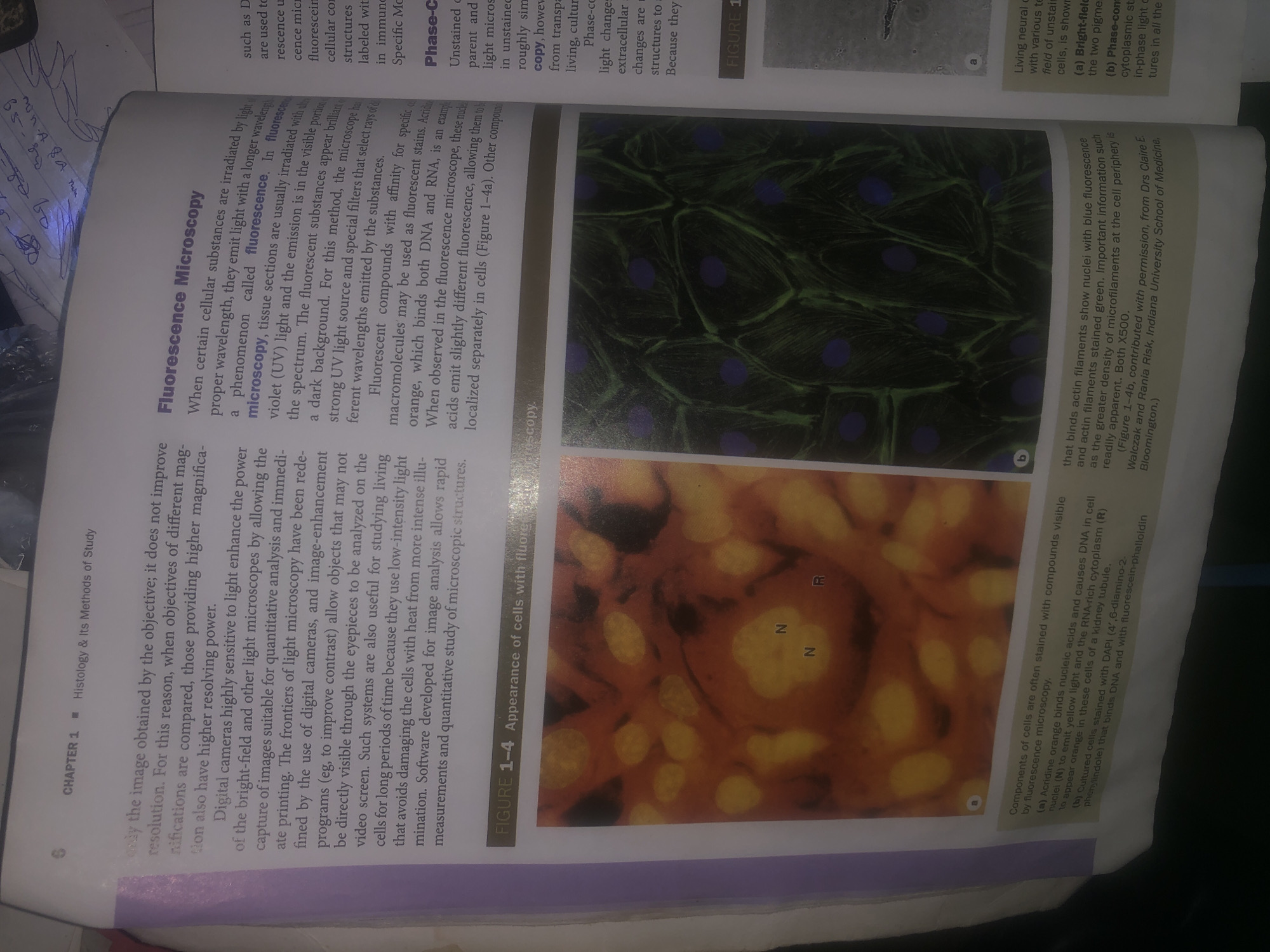
The image displays two figures, labeled 1-4a and 1-4b, illustrating cells as viewed under a fluorescence microscope. Figure 1-4a shows cells with yellow-fluorescing nuclei (labeled 'N') and cytoplasm (labeled 'R'). Figure 1-4b depicts cells with blue-fluorescing nuclei and green-fluorescing actin filaments. The text describes the principles and applications of fluorescence microscopy, detailing how certain cellular components are stained with fluorescent compounds and visualized under specific light wavelengths. The image and its accompanying text are from a textbook chapter on Histology & Its Methods of Study. There is no information in the image or text that relates to Wukari, Nigeria.
Bigstev

Jun 12, 2026, 1:31 AM

Wukari, Nigeria

Moment

Stake attention in this memory

curious
educational
analytical
informative
scientific

The image displays two figures, labeled 1-4a and 1-4b, illustrating cells as viewed under a fluorescence microscope. Figure 1-4a shows cells with yellow-fluorescing nuclei (labeled 'N') and cytoplasm (labeled 'R'). Figure 1-4b depicts cells with blue-fluorescing nuclei and green-fluorescing actin filaments. The text describes the principles and applications of fluorescence microscopy, detailing how certain cellular components are stained with fluorescent compounds and visualized under specific light wavelengths. The image and its accompanying text are from a textbook chapter on Histology & Its Methods of Study. There is no information in the image or text that relates to Wukari, Nigeria.

Loading AttnAds…

transactions
revenues
stakers
Earliest
Latest
Highest stake

No transactions found

More from this user

More from Wukari

Bigstev

Jun 12, 2026, 1:31 AM

Wukari, Nigeria

Licensed under Creative Commons Attribution 4.0 International License. You are free to share and adapt this content with proper attribution.