
Sign in
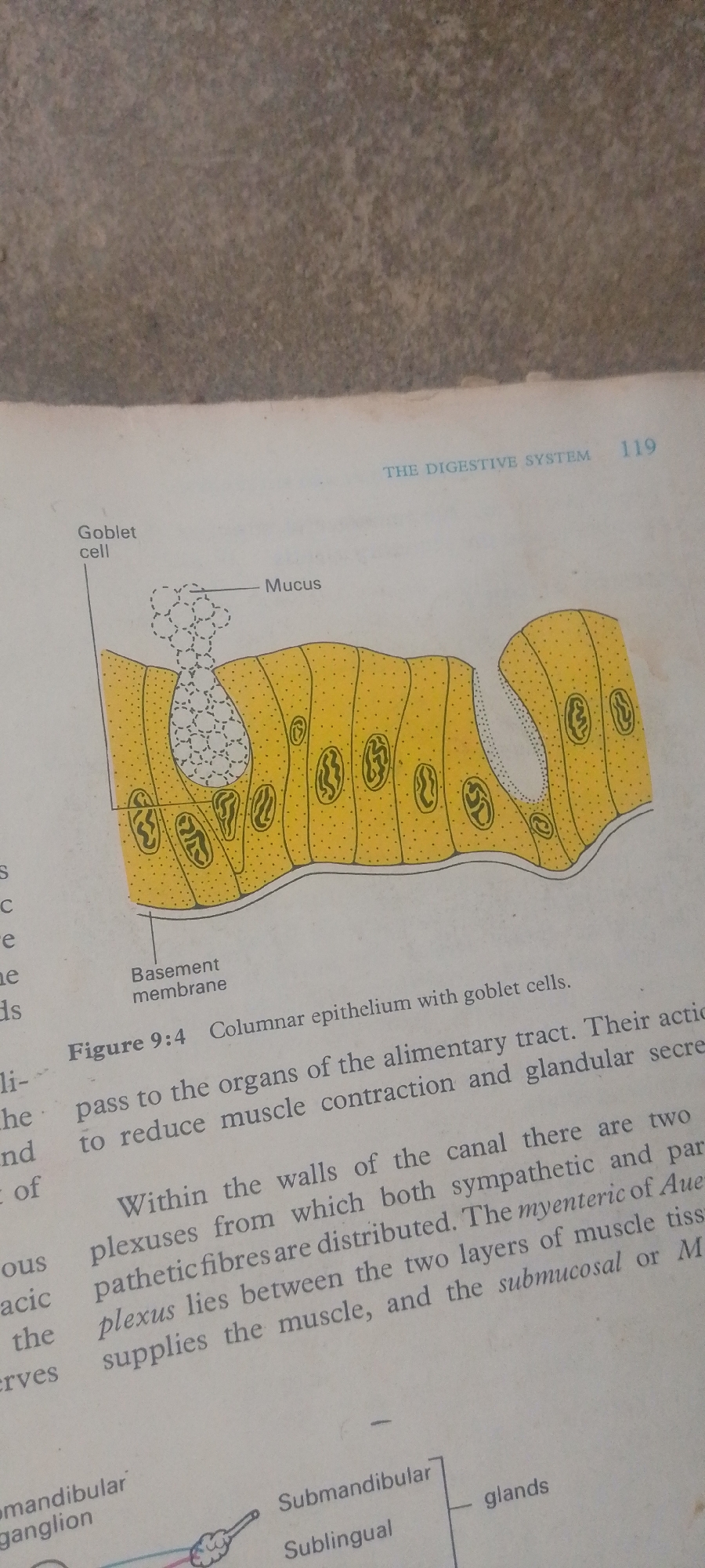
The image is a photograph of a page from a biology textbook. The page contains an illustration of columnar epithelium with goblet cells, a diagram related to glands, and accompanying text.

The illustration on the top half of the page depicts a cross-section of the lining of an organ, likely part of the digestive system, as indicated by the text "THE DIGESTIVE SYSTEM". It shows tall, columnar cells, some of which are goblet cells filled with mucus. A label points to "Mucus" being secreted by a goblet cell. Below the epithelium, a "Basement membrane" is labeled.

The text below the illustration provides a caption for Figure 9:4, stating "Columnar epithelium with goblet cells." Further text describes the function of these cells and their distribution within the alimentary tract, mentioning sympathetic and parasympathetic nerve plexuses.

The bottom portion of the page shows a partial diagram of glands, with labels for "Submandibular," "Sublingual," and "glands." There is also a label for "mandibular ganglion."

The overall scene is that of an educational resource, with clear diagrams and explanatory text designed to teach about biological structures and functions. There are no people, specific time of day, weather, or emotional cues visible. The location context provided (Jauro Yinu, Nigeria) is external information and not derivable from the image itself. The visible text is entirely scientific and instructional in nature, including labels for anatomical features and page numbering ("119").
FM-2L6Lq2

Jun 30, 2026, 5:10 PM

Jauro Yinu, Nigeria

Moment

Stake attention in this memory

academic
analytical
studious
curious
objective

The image is a photograph of a page from a biology textbook. The page contains an illustration of columnar epithelium with goblet cells, a diagram related to glands, and accompanying text. The illustration on the top half of the page depicts a cross-section of the lining of an organ, likely part of the digestive system, as indicated by the text "THE DIGESTIVE SYSTEM". It shows tall, columnar cells, some of which are goblet cells filled with mucus. A label points to "Mucus" being secreted by a goblet cell. Below the epithelium, a "Basement membrane" is labeled. The text below the illustration provides a caption for Figure 9:4, stating "Columnar epithelium with goblet cells." Further text describes the function of these cells and their distribution within the alimentary tract, mentioning sympathetic and parasympathetic nerve plexuses. The bottom portion of the page shows a partial diagram of glands, with labels for "Submandibular," "Sublingual," and "glands." There is also a label for "mandibular ganglion." The overall scene is that of an educational resource, with clear diagrams and explanatory text designed to teach about biological structures and functions. There are no people, specific time of day, weather, or emotional cues visible. The location context provided (Jauro Yinu, Nigeria) is external information and not derivable from the image itself. The visible text is entirely scientific and instructional in nature, including labels for anatomical features and page numbering ("119").

Symbol

FC929

Volume

3,268

Creator

+$0.00

Revenue

+$0.00

TVL

$0.28

1D
1W
1M
All

Loading AttnAds…

transactions
revenues
stakers
Earliest
Latest
Highest stake

1

FM-FSRgn1

Staked 16d ago

$0.28 USD

+$0.00 USD

+0.5%

2

Rev Bot 🤖💰

Injected revenue 16d ago

Revenue bonus for the last stake.

+$0.00 USD

More from this user

More from Jauro Yinu

FM-2L6Lq2

Jun 30, 2026, 5:10 PM

Jauro Yinu, Nigeria

Licensed under Creative Commons Attribution 4.0 International License. You are free to share and adapt this content with proper attribution.