Stake attention in this memory
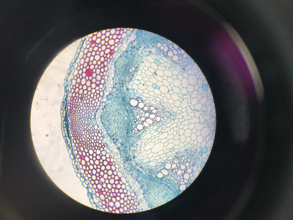
This image displays a circular, magnified view of a stained transverse section of a plant stem, observed through an optical microscope. The specimen exhibits distinct cellular tissues. An outermost compact epidermal layer is visible, followed by cortical parenchyma cells, primarily stained blue, and includes scattered smaller, round cells stained pink. Vascular bundles, characterized by larger, blue-stained xylem vessels and smaller, densely packed phloem cells, are arranged in a ring. The central pith region comprises large, blue-stained parenchyma cells. The area immediately surrounding the circular microscopic field is uniformly dark. No individuals, actions, or interactions are depicted within the frame. This scene is located in Midrand, South Africa.
No transactions found