
Sign in
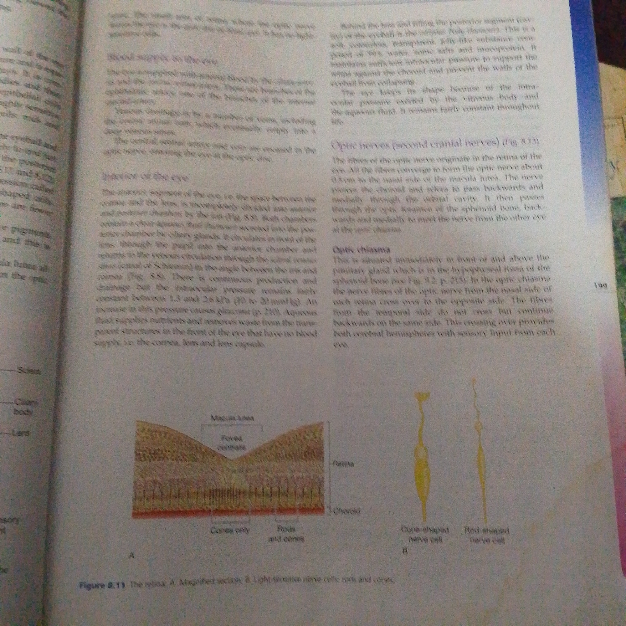
The image is a scan of a page from a biology textbook. The text discusses the anatomy and physiology of the eye, specifically focusing on blood supply, the interior of the eye, optic nerves, and the optic chiasma. There are two diagrams. Diagram A is a magnified cross-section of the retina, showing the macula lutea, fovea centralis, and the layers containing cones and rods. Diagram B shows two diagrams of photoreceptor cells: a cone-shaped nerve cell and a rod-shaped nerve cell. The text and diagrams are printed in black and white. The image itself is slightly blurry and shows the texture of the paper. The bottom of the page has a page number "199". There is no information in the image that relates to the city of Jalingo, Nigeria.
FM-66btC3

Jun 30, 2026, 1:24 PM

Jalingo, Nigeria

Stake attention in this memory

informative
educational
scientific
detailed
visual

The image is a scan of a page from a biology textbook. The text discusses the anatomy and physiology of the eye, specifically focusing on blood supply, the interior of the eye, optic nerves, and the optic chiasma. There are two diagrams. Diagram A is a magnified cross-section of the retina, showing the macula lutea, fovea centralis, and the layers containing cones and rods. Diagram B shows two diagrams of photoreceptor cells: a cone-shaped nerve cell and a rod-shaped nerve cell. The text and diagrams are printed in black and white. The image itself is slightly blurry and shows the texture of the paper. The bottom of the page has a page number "199". There is no information in the image that relates to the city of Jalingo, Nigeria.

Loading AttnAds…

transactions
revenues
stakers
Earliest
Latest
Highest stake

No transactions found

More from this user

More from Jalingo

FM-66btC3

Jun 30, 2026, 1:24 PM

Jalingo, Nigeria

Licensed under Creative Commons Attribution 4.0 International License. You are free to share and adapt this content with proper attribution.